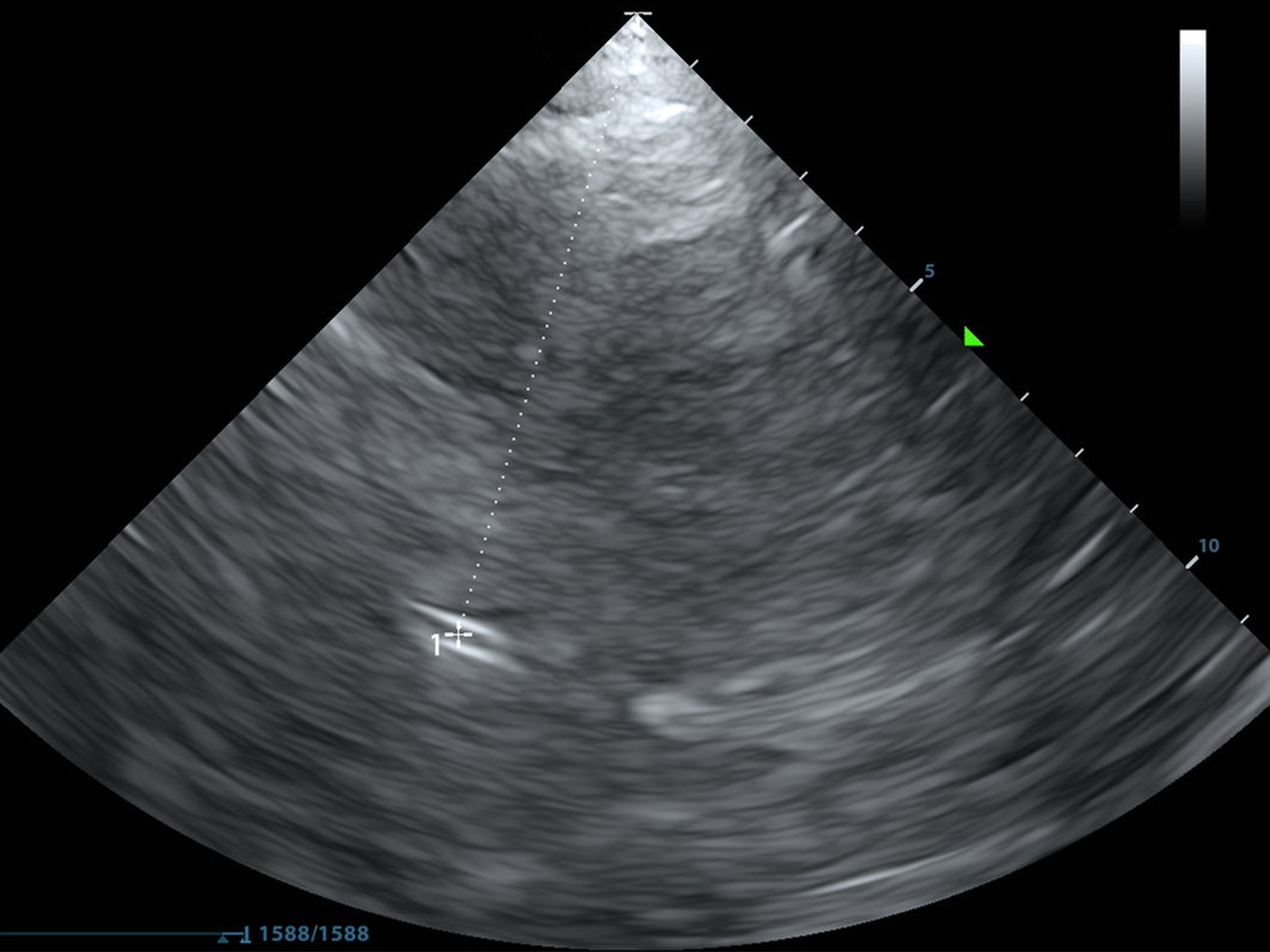
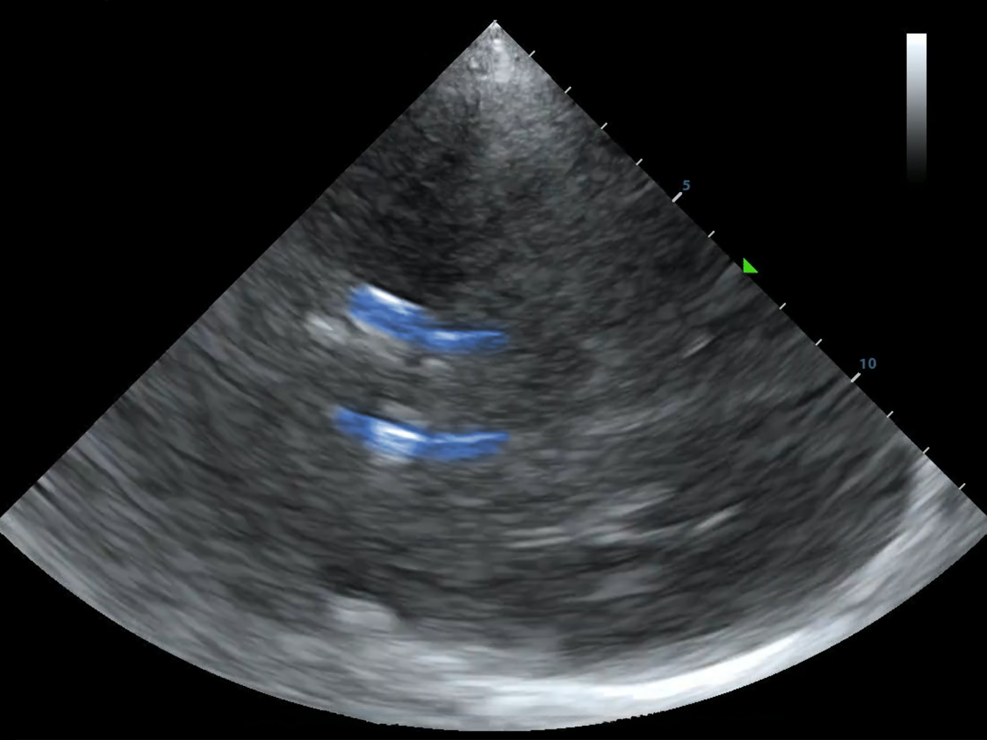
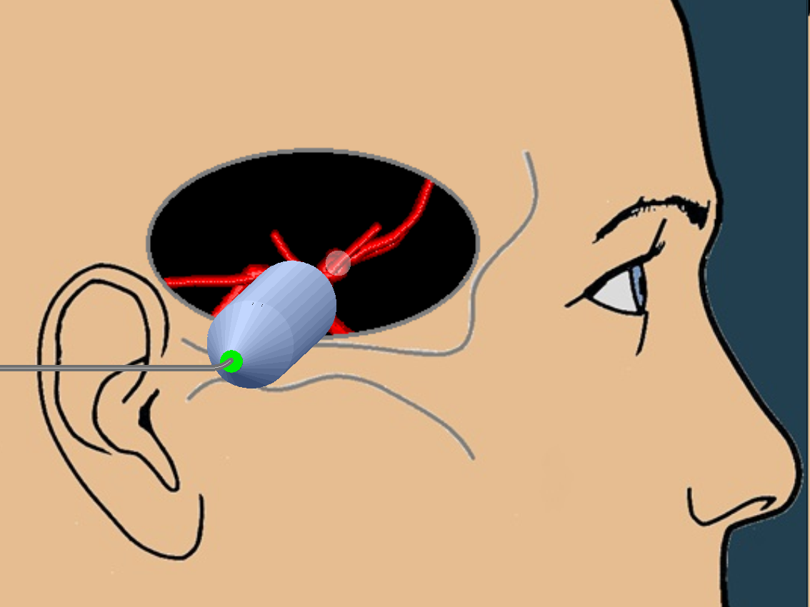
This is the circle of Willis. As you now know, it's found between the mesencephalon and diencephalon planes, with the probe angled about 10 degrees cephalad from perpendicular to the temporal bone. If you have difficulty with these views, go back and read "The Basics" first!
Notice that the MCA is reaching right out at you.
Also notice that if your color box is too small you might mistake the PCA or intracranial ICA for the MCA...
Once you've got a good juicy MCA in view. Turn on Pulse Wave (PW) doopler and put the gate--the little "=" sign--on the proximal MCA, just after it branches off of the ICA and ACA.
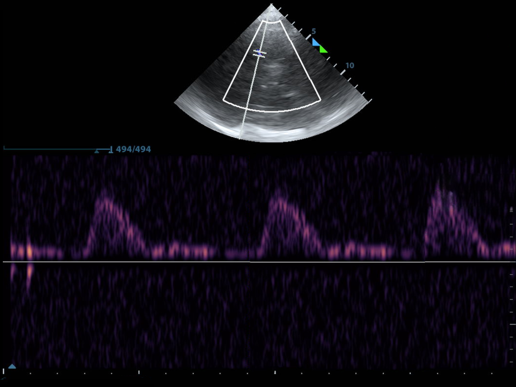
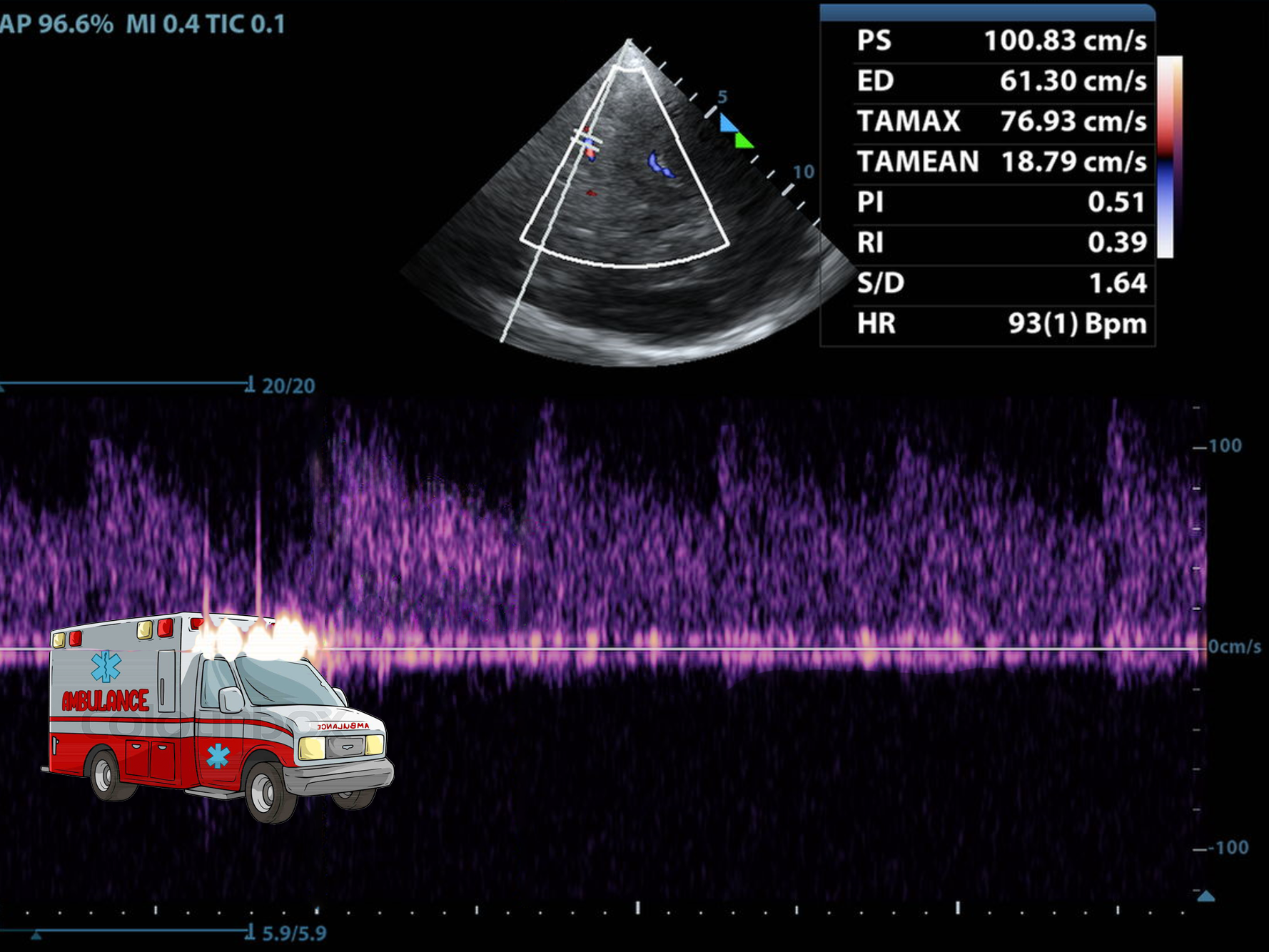
You should get a waveform that looks something like this:
The flow sounds a lot like Owen Wilson saying, "wow."
Check it out!
The shape of the waveform alone is useful information. Normal cerebral blood flow has constant forward flow. That is, there's forward flow in diastole, too. It's the reason you don't pee your pants and forget who you are in between heartbeats.
Good diastolic flow should be 30-50% the height of the systolic peak. The waveform looks like a bunch of sneakers lined up.
With blunted diastolic flow, the diastolic portion is depressed. Mildly depressed diastolic flow can be seen in patients with chronic microvascular disease and in mildly increased ICP. Significantly blunted diastolic flow, as below, should raise the suspicion for dangerously elevated ICP if present bilaterally.
The waveform looks more like heels than sneakers. This can be a periherniation waveform.
When the diastolic portion of the waveform shows retrograde flow, or flow below the baseline, the waveform looks like a boot heel stomping through the floor.
There should be not booting, scooting, and/or boogieing in the cerebrovascular system.
This is a periherniation waveform if seen bilaterally and should be intervened upon. Before you leave the room. It is unlikely to see waveform before a patient completely decompensates.
Below is an MCA waveform in a patient with a large basal ganglia bleed and altered level of consciousness shortly before neurosurgery placed and External Ventricular Drain (EVD):
More high heels than sneakers, right?
Below is an MCA/distal ICA waveform in a patient with a ruptured aneurysm shortly after decompensation and intubation. After seeing this waveform, neurointensive care administered hypertonic saline and neurosurgery was called to bedside and place an EVD prior to CT imaging:
